Oral and Maxillofacial Radiology
Dental radiology, panoramic X-rays and diagnostic imaging for individual dental treatment planning at Neovadent in Kırklareli, Turkey.
Oral and Maxillofacial Radiology
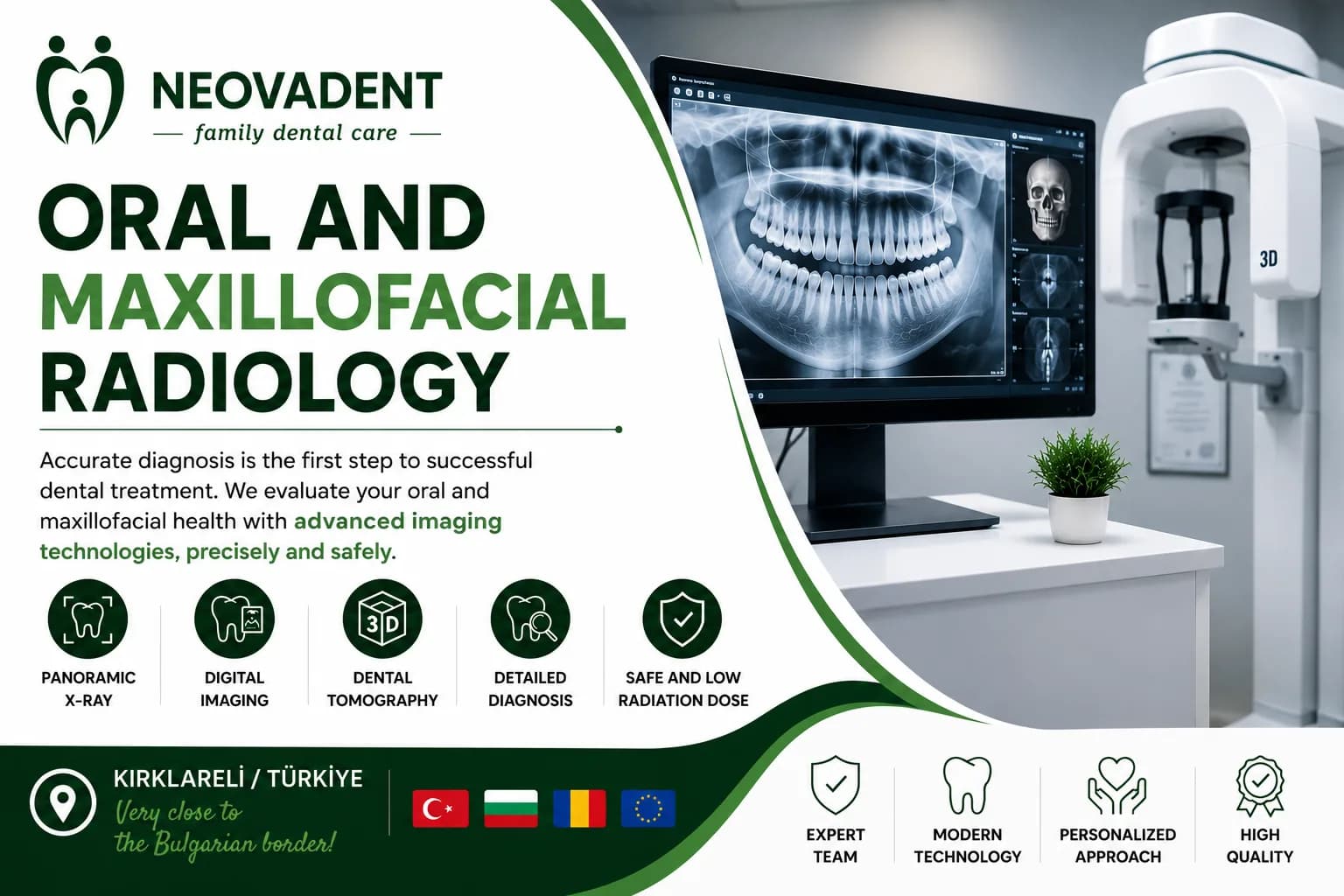
Oral and maxillofacial radiology is a field of dentistry concerned with the clinical and diagnostic imaging assessment of conditions affecting the teeth, jawbones and surrounding tissues.
Accurate diagnosis is one of the fundamental stages of appropriate dental treatment planning. Some conditions may not be detected through visual examination alone, and diagnostic imaging may be required where clinically indicated.
At Neovadent Dental Clinic in Kırklareli, clinical findings and necessary diagnostic images are evaluated together to develop an individual treatment plan.
What Does Oral and Maxillofacial Radiology Include?
Diagnostic imaging may assist in evaluating:
Dental cavities
Tooth roots and surrounding tissues
The jawbone
Impacted and partially impacted teeth
The position of wisdom teeth
Bone structure before dental implant treatment
Cysts and other changes
Orthodontic treatment planning
Root canal treatment
Anatomical structures within the maxillofacial region
The appropriate imaging method is determined according to the patient's individual clinical needs.
What Is a Panoramic Dental X-Ray?
A panoramic dental X-ray provides an overall image of the upper and lower teeth, jawbones and certain surrounding anatomical structures.
It may be used when evaluating impacted wisdom teeth, planning dental implant treatment, conducting orthodontic assessments and examining overall oral health.
Not every patient requires a panoramic X-ray. The need for diagnostic imaging is determined according to clinical findings.
When May Dental Tomography Be Required?
Three-dimensional dental imaging may provide more detailed information about the jawbone and surrounding anatomical structures.
It may be considered in certain cases involving dental implant planning, impacted teeth, bone assessment or complex oral surgical procedures.
The appropriate imaging method should be selected according to clinical necessity.
Radiological Assessment Before Dental Implant Treatment
Before dental implant placement, evaluating the bone structure in the intended implant area is an important part of treatment planning.
Diagnostic imaging may help assess available bone height and width, nearby anatomical structures and other factors that could affect implant placement.
The required imaging method may differ from one patient to another.
Radiological Assessment of Impacted Wisdom Teeth
Wisdom teeth may remain fully or partially impacted within the jawbone. Diagnostic imaging can help evaluate the position of the tooth and its relationship with neighbouring teeth and nearby anatomical structures.
This information may assist in determining whether observation or surgical treatment should be considered.
Why Is Accurate Dental Diagnosis Important?
Many oral health problems may not cause noticeable symptoms during their early stages. Combining clinical examination with appropriate diagnostic imaging can provide a more comprehensive understanding of a patient's oral health.
The aim is to establish an appropriate diagnosis and create a treatment plan based on the patient's actual clinical needs.
Dental Radiology at Neovadent in Kırklareli, Turkey
Neovadent Dental Clinic is located in Kırklareli, in the European part of Turkey and close to the Bulgarian border. Clinical examinations and necessary diagnostic imaging are important parts of our individual dental treatment planning process.
Accurate diagnosis supports the planning of different dental treatments, including implant dentistry, oral surgery, endodontics and orthodontics.
Contact Neovadent to learn more or schedule a consultation and individual dental assessment.
Frequently Asked Questions
1. What is oral and maxillofacial radiology?
It is a field of dentistry that uses clinical examination and appropriate diagnostic imaging methods to evaluate the teeth, jawbones and surrounding tissues.
2. Is an X-ray necessary at every dental examination?
No. The need for an X-ray depends on the patient's symptoms, clinical findings and planned treatment.
3. What does a panoramic dental X-ray show?
It provides an overall image of the teeth, upper and lower jawbones, impacted teeth and certain surrounding anatomical structures.
4. Is diagnostic imaging necessary before dental implant treatment?
An assessment of the bone structure and surrounding anatomical features is generally required before implant treatment. The specific imaging method is selected according to individual needs.
5. Can an impacted wisdom tooth be seen on an X-ray?
Yes. Diagnostic imaging can help determine the tooth's position and its relationship with neighbouring teeth and surrounding anatomical structures.
6. What is the difference between a panoramic X-ray and dental tomography?
A panoramic X-ray provides a general two-dimensional image, while dental tomography may offer more detailed three-dimensional information about a specific area.
7. Are dental X-rays safe?
Dental imaging should be performed when clinically indicated, using appropriate techniques and with the aim of keeping radiation exposure as low as reasonably achievable.
8. Is an X-ray alone enough to make a diagnosis?
Not always. Diagnostic images are evaluated together with the patient's symptoms, medical history and clinical examination findings.
İç link önerileri: English Dental Implants, Oral and Maxillofacial Surgery, Wisdom Tooth Removal, Root Canal Treatment and Orthodontics pages.
Other services
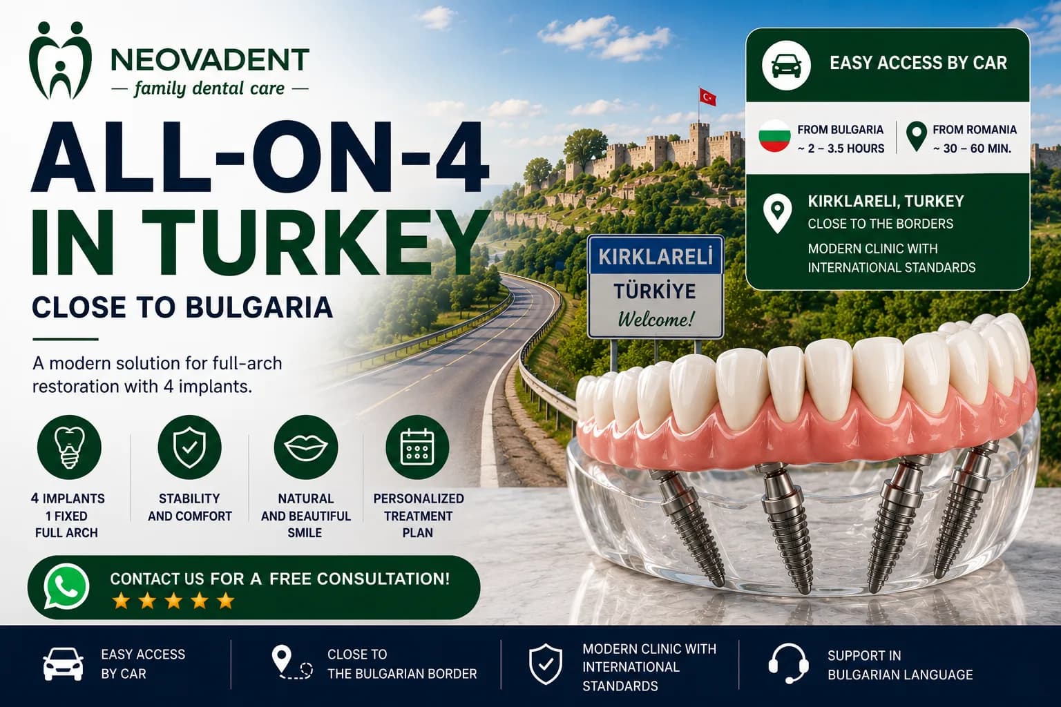
All-on-4 in Turkey | Near Bulgaria & Romania | Neovadent
Discover All-on-4 treatment in Turkey at Neovadent, Kırklareli. Modern digital dentistry, personalized treatment plans, and convenient access from Bulgaria and Romania.

Dental Implants in Turkey Near Bulgaria | Neovadent
Looking for dental implants in Turkey? Neovadent offers modern implant treatments in Kırklareli with convenient access from Bulgaria and Romania.
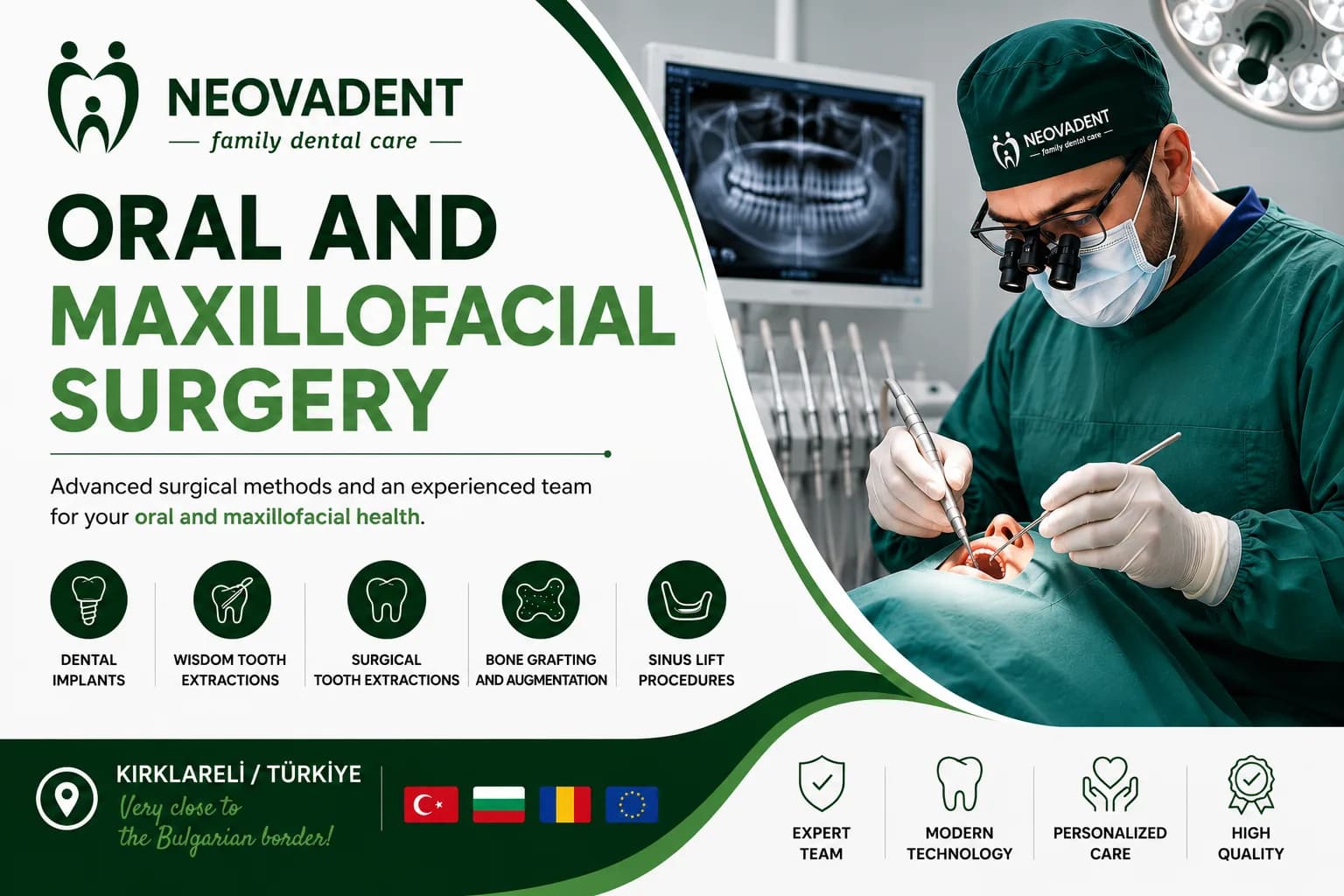
Oral and Maxillofacial Surgery
Oral surgery, dental implants and impacted wisdom tooth removal at Neovadent in Kırklareli, Turkey. Individual treatment planning for every patient.